Thoracic ultrasound is mostly utilized to locate a target organ- or disease-specific condition and is often used as a complement to other imaging, such as chest radiograph, computed tomogram, or magnetic resonance imaging.
In this blog, Manhattan pulmonologist Dr. Marc Bowen will explain the top reasons for getting a thoracic ultrasound.
What is a thoracic ultrasound?
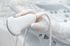
A thoracic or pulmonary ultrasound is a non-invasive diagnostic test that uses a transducer (wand-like device) on the outside of the skin, which utilizes sound waves to produce an image of the area being examined. It’s used to assess the structures and organs within the chest, such as the lungs and pleural space (the area between the lungs and the chest’s interior wall). It can also be used to examine the mediastinum (the area of the chest that contains the heart, esophagus, lymph nodes, and other structures).
An ultrasound provides a doctor with a quick visualization of what’s occurring within this space, all from outside the body.
What are the top reasons for getting a thoracic ultrasound?
A pulmonologist may recommend this procedure if you’re experiencing the following symptoms or conditions:
- Symptoms of pneumonia, such as a cough, chills, fatigue, and fast, shallow breathing
- Pulmonary edema (excess water in the lungs), which can cause chest pain, shortness of breath when you lie down, fast or shallow breathing, or wheezing
- Pleural effusion (fluid around the lungs), which can cause chest pain that can be sharp, fast breathing or shortness of breath, or coughing
- Pneumothorax (collapse of the lung), which can cause fast or shallow breathing, shortness of breath, chest tightness, or coughing
What’s involved with a thoracic ultrasound?
During this procedure, gel will be placed on a special device called a transducer, as well as on the skin in the area to be examined. This allows the transducer to move smoothly over the skin, providing the best visualization of what’s going on inside your body.
The transducer emits ultrasound waves that move through the body to the organs, tissues, and other interior structures. The waves bounce off these structures and transmit information back to the transducer.
This information is then converted into an image that the doctor can view on a monitor to examine what’s occurring within your chest.
What are the benefits of a thoracic ultrasound?
Benefits of this procedure include:
- Performed in-office, making it quick and convenient for you
- Provides accurate results in real time, which helps the doctor make a quick, accurate diagnosis
- Doesn’t use radiation
- Doesn’t use contrast dye, so there’s no need to drink a bad-tasting liquid or have an injection
- Avoids possible allergic reactions that some people have with contrast dye
- Can be performed during pregnancy
Where can I get a thoracic ultrasound performed in NYC?
Dr. Bowen is a New York City pulmonologist who is dedicated to providing the highest level of personalized patient experience. He has nearly three decades of clinical practice specializing in Pulmonary Disease, Critical Care Medicine, and Internal Medicine.
After undergoing a pulmonary or thoracic ultrasound, Dr. Bowen will immediately be able to make an accurate diagnosis within a matter of minutes. This enables you to start any needed treatment as quickly as possible.
After making a diagnosis, Dr. Bowen will explain your treatment options, answer any questions you may have, and discuss the next steps for treating your condition and restoring normal breathing function.
If you’re suffering from shortness of breath, chronic cough, a tight feeling in your chest, or sharp chest pain, please contact our office to schedule an evaluation so Dr. Bowen can determine whether you’ll benefit from a pulmonary or thoracic ultrasound. We offer same-day appointments for your convenience.

